About MEG
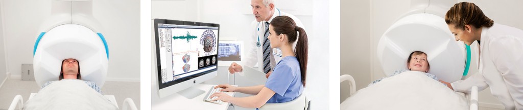
Magnetoencephalography (MEG) is a functional brain imaging technology used to record the magnetic fields associated with electric currents generated by synchronously active populations of neurons in the brain. It uniquely combines millisecond temporal resolution with millimeter spatial resolution. Therefore, it is the only non-invasive technique which can measure the fastest brain events or rhythms, and then accurately localize them. When combined with anatomical images from MRI, it can help guide surgeries and reveal the most complex workings of the human brain. It is completely safe, silent and non-intimidating.
MEG was originally developed by David Cohen at the Massachusetts Institute of Technology in the 1970s and is based on the use of highly sensitive detectors called SQUIDs, or super-conducting quantum interference devices. These detectors can accurately measure the occurrence of spontaneous or evoked brain activity. The most important clinical application is presurgical planning for patients suffering from intractable epilepsy. MEG detects the signature “spikes” which can indicate the existence and location of the onset of epileptic activity.